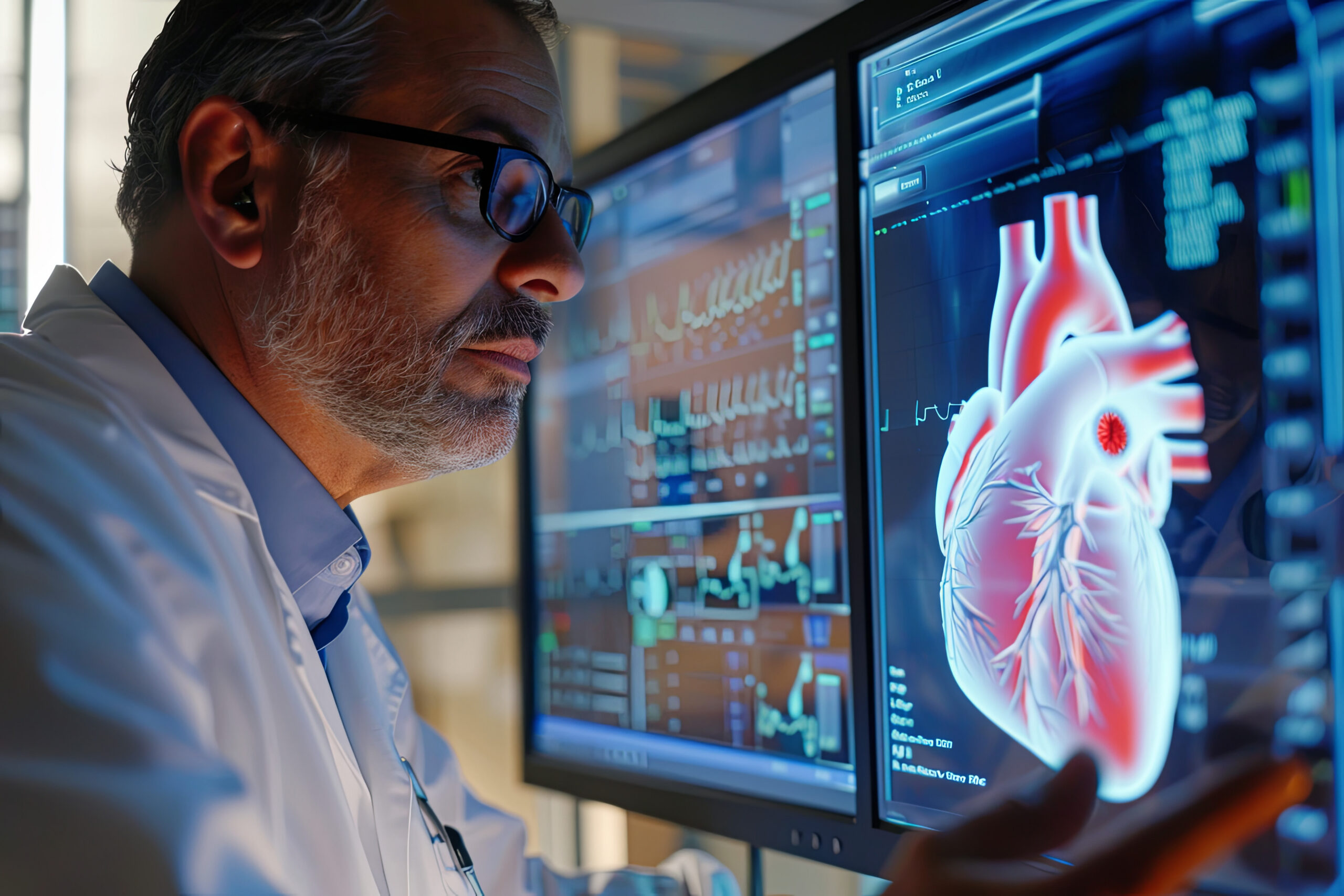
The image of good health
State-of-the-art Imaging Centers.
We offer the most advanced imaging technology available to accurately diagnose heart disease and meet the individualized needs of every cardiac patient. Our imaging centers at our Arville, North Tenaya, and North Pecos clinics are the only in Southern Nevada that are owned and operated by a private cardiology practice.

Non-invasive
Low radiation
Highly accurate
Board certified nuclear cardiologist
Coronary calcium score
This simple 5-minute test can help diagnose possible heart disease.
A Coronary Calcium Score is a specialized test that uses x-rays to create images of the heart and detect the presence of calcium containing plaque in the arteries supplying the heart muscle. This 5-minute test requires no special preparation and has a strong ability to reclassify cardiovascular risk. A calcium score of 0 is associated with low risk of adverse cardiovascular events. Higher calcium scores can signal higher risks, including heart attack or the need for a coronary stent. In individual cases with borderline or uncertain risk, personal concerns or statin side effects, this is one of the best tools we have today to guide heart management and treatment plans.
CTA scan
Stress TestCTA stands for CT Angiography. This specialized scan focuses particularly on the heart’s blood vessels.
These images of the heart provide detailed pictures of the blood vessels that supply the heart muscle, also known as the coronary arteries. If these arteries are blocked and left untreated, a heart attack can occur.
How does it work?
The coronary CT Angiogram takes images of the heart arteries that are clear enough for any blockages to be seen. It can be used to evaluate a patient with chest pain or to clarify an uncertain stress test. It is also an excellent tool for assessing patients with symptoms of numerous other conditions, including pulmonary emboli (blood clots in the lungs) and aortic aneurysm (a bulge or ballooning in the wall of the main artery carrying blood from the heart).
What can the CTA rule out?
The CT Angiography is consistently able to rule out the significant narrowing of the major coronary arteries. It can also non-invasively evaluate for plaque in the artery walls – a condition that can lead to a serious blockage if left untreated. This advanced warning can allow a patient to seek medical treatment and/or make lifestyle changes that may prevent a future heart attack.
Revolutionizing the diagnosis and treatment of Peripheral Arterial Disease (PAD)
Peripheral Arterial Disease (PAD) is generally associated with blocked arteries in the legs. The blockage is most often the result of a chronic buildup of hard fatty material within the inside lining of the arterial wall of the legs. This ultimately narrows and blocks the flow of blood carrying oxygen and nutrients to the limb. The femoral and popliteal arteries are the major arterial blood supply to the lower extremities and are a common location for atherosclerotic disease to develop.
Using the CTA to diagnose PAD allows your doctor to more easily visualize blood flow through the lining of artery walls. This state-of-the-art test provides your medical team with the ability to properly non-invasively diagnose or rule out PAD.
A safer alternative than the Angiogram
The CTA is a much safer diagnostic tool than the more invasive angiogram, which requires a surgical setting where a catheter is placed in your heart. The CTA is a completely non-invasive heart scan, avoiding the complications often associated with the traditional angiogram.
PET scan
PET stands for Positron Emission Tomography and is the most advanced and accurate stress testing technology for visualizing blood flow through the heart arteries to the heart muscle. PET can be an alternative to other more invasive tests. A cardiac PET scan is a type of stress test in which blood flow to the heart muscle is visualized with a radiotracer and a special scanner. PET provides an accurate, non-invasive assessment of the coronary arteries. Pictures are taken to determine if blood flow is restricted by a narrowing of the coronary arteries.
How is a PET Scan different from other tests?
Clinical studies have shown that PET scans are more accurate than other widely used nuclear stress tests. These other tests are associated with significant “false positive” results. False positives are results that show coronary heart disease where none exists and can lead to a patient having to undergo an unnecessary procedure. Because PET scans are so accurate, they are often used to confirm other tests if a false positive is suspected. PET scans also avoid the false negatives that are common with other tests and can lead to an unexpected heart attack.
Dual source CT
Bringing advanced CT to patients for the first time
More than 55 million computed tomography (CT) exams are performed in the U.S. each year, enabling physicians to obtain detailed three-dimensional images of a patient’s anatomy to pinpoint disease earlier, with greater precision, and without surgery. Until now, CT clarity has been measured in “slices” – cross-sectional horizontal and vertical images of a patient’s body – with advanced CT scanners producing 64 slices.
Physicians at Nevada Heart and Vascular Imaging Center are able to go beyond slices with the advanced technology of SOMATOM Definition. The dual-source CT system has twice the speed and resolution of even the most advanced single-source CT system, offering capabilities not available through any other diagnostic imaging technology. Our physicians can bring the benefits of advanced CT to nearly any patient – regardless of size, condition or heart rate – using the SOMATOM Definition.
Echocardiogram
An echocardiogram (echo) is a simple imaging test that helps your doctor evaluate your heart. This test is safe and painless, and can be performed in a hospital, testing center, or doctor’s office. The echo bounces harmless sound waves (ultrasound) off the heart. A transducer – a device that looks like a microphone – is used to help show the size of your heart, as well as revealing the health of the heart’s chambers and valves.
Before your echo
- Discuss any questions or concerns you have with your doctor.
- Mention all over-the-counter or prescription medications, herbs, or supplements you are taking.
- Allow extra time for checking in.
- Wear a two-piece outfit for the test. You may be asked to remove clothing and jewelry from the waist up. If so, you’ll be given a short hospital gown.
During your echo
Most echo tests take 10-20 minutes. Small pads (electrodes) are placed on your chest to monitor your heartbeat. A transducer coated with cool gel is moved firmly over your chest, creating the sound waves that make images of your heart. At times, you may be asked to exhale and hold your breath for a few seconds, as air in your lungs can affect the images. The transducer may also be used to do a Doppler study, an additional test measuring the direction and speed of blood flowing through the heart. During the test, you may hear a “whooshing” sound, which is simply the sound of blood flowing through the heart. The images of your heart are stored on a computer or recorded on video, where your doctor can review them later.
After your echo
Unless your healthcare provider tells you otherwise, you can return to normal activity. Be sure to keep all follow-up appointments.
Your test results
Your doctor will discuss your test results with you during a future office visit. The results help your doctor plan your treatment and schedule any other tests that are needed.
Nuclear imaging
Cardiac nuclear imaging is also called a “perfusion scan.” A tracer (a small amount of safe radioactive matter) is delivered into the bloodstream. A camera then scans the tracer in the blood as it flows through the heart muscle. This test can be performed in a hospital or testing center, and the tracer safely leaves your body within hours.
Before your test
The entire test will take a few hours. For best results, prepare for your test as directed. When you schedule your test, be sure to mention all medications you take, and ask if you should stop taking any of them the day of the test. Stop smoking and avoid caffeine for as long as directed. Sips of water are permitted, but don’t eat or drink for 4-6 hours before the test. On the day of the test, dress for comfort and wear a two-piece outfit, top and bottoms. Be sure to wear walking shoes.
During your test
You may be asked to change into a hospital gown for the test. Picture scans may be taken while you rest before exercising, or these resting scans may need to take place on a return visit later the same day, or the next day. You will be attached to EKG and blood pressure monitors, and an IV (intravenous) line will be started in your arm. You will then exercise on a treadmill or stationary bike for a few minutes to increase the rate of blood flow to your heart muscle. Speak up when you feel you cannot exercise for even one more minute. At this point, the tracer is given to you through the IV. For those who cannot exercise, special medications can be used to increase heart rate. After you have received the tracer, you will be positioned on the scanning bed, where you must lie very still for up to 30 minutes. During this time, a scanning camera will be taking images showing where blood flows through your heart muscle.
After your test
Before going home, ask when you may eat and when to resume taking any medications you were told to avoid on your testing day. If you need to return for resting scans, be sure to follow any instructions. Most patients can go back to their normal routine as soon as all parts of the test are finished.
Report any symptoms
Be sure to tell the doctor if you feel any of the following during the test:
- Chest, arm, or jaw discomfort
- Severe shortness of breath
- Dizziness or lightheadedness
- Leg cramps or pain
Let the technologist know:
- Any medicines you take
- If you have diabetes, knee or hip problems, arthritis, asthma, or chronic lung disease
- If you have had a stroke or have vascular disease of the leg
- If you are pregnant, think you might be, or are nursing
Vascular ultrasound
What is a vascular ultrasound study?
This test examines the blood flow in the major arteries and veins in the body with the use of ultrasound (high-frequency sound waves). A vascular ultrasound study is ordered to diagnose blockages in a blood vessel that might cause a variety of symptoms or problems.
This procedure uses sound waves to obtain images and measure the blood flow. These images are analyzed to determine whether you have blockages in your arteries, blood clots in your veins, or if an aneurysm is present.
An instrument that transmits high-frequency sound waves called a transducer is placed over an artery or vein. The transducer receives the echoes of the sound waves and transmits them as electrical impulses. The ultrasound machine converts these impulses into images that are recorded by the technologist.
VNUS closure procedure
During the VNUS Closure™ procedure, the patient is placed under oral sedation. A small, sterile catheter is inserted into the diseased vein. Using radiofrequency ablation, the catheter quickly heats the vein, causing it to collapse and seal shut. The blood flow is redirected to the remaining healthy veins. The procedure is performed in segments and is usually completed in just a few minutes.
Following the procedure, a bandage is placed on the insertion site, and a compression stocking is used to aid healing. You may be encouraged to refrain from strenuous activity for a time. Most patients report minimal to no scarring, bruising or swelling, and can resume normal activities within one day.
A study on post-closure patients who remained free of venous reflux for one year revealed that 92% were still reflux-free after five years.
The Patient Portal that does it all.
Schedule your appointment. Message your care team. View test results, pay bills, and more. With your secure password, you can log in 24 hours a day, 7 days a week from the comfort and privacy of your home or office. Getting started is simple.
